Overcoming Limitations in Multimodal Microscopy for Bacterial Spore Germination
Phase contrast microscopy is a widely used technique for visualising transparent specimens without staining, but its conventional setup can present significant challenges in experimental configurations.
Specifically, the bulky components required for phase contrast often restrict access to the sample area on an inverted microscope frame, making it difficult to integrate additional techniques like Scanning Ion Conductance Microscopy (SICM).
SICM uses an electrode positioned just above the sample surface to map topography without physical contact, a critical requirement for certain advanced imaging applications.
Dr. Gabriel Meloni at Warwick University sought to combine SICM with phase contrast microscopy to monitor bacterial spore germination. However, traditional phase contrast assemblies on inverted microscopes presented a dual problem: the components impeded the precise positioning of the SICM electrode tip, and the electrode assembly obstructed light from the transmitted light source, compromising image quality.
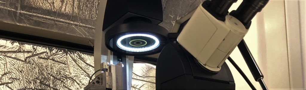
Cairn Research's Aura Illuminator—Enhancing Multimodal Microscopy Capabilities
To address these challenges, Cairn Research provided a bespoke solution with the Aura Illuminator, a novel phase contrast lighting system that streamlines the traditional configuration by reducing the number of components. The Aura Illuminator eliminates the need for a conventional condenser, freeing up critical space above the sample to accommodate the SICM electrode assembly.
The Aura Illuminator features concentric LED rings tailored to match PhL, Ph1, and Ph2 phase contrast objectives, providing flexibility and precision in phase contrast imaging.
Cairn Research also developed a custom adapter, enabling the Aura Illuminator to integrate seamlessly with the transmitted illumination pillar of Dr. Meloni’s existing Leica DMI4000B microscope.
This tailored approach allowed for a compact and efficient setup, facilitating the simultaneous use of SICM and phase contrast microscopy.
Expanded Functionality and Enhanced Imaging Performance
By partnering with Cairn Research, Dr. Meloni transformed an under-utilised microscope into a powerful multimodal imaging platform capable of both SICM and phase contrast microscopy.
This integration allowed for the detailed study of bacterial spore germination, demonstrating the effectiveness of Cairn Research’s innovative solutions in overcoming technical barriers in microscopy.
Special recognition goes to Kevin Webb from Nottingham University, who conceptualised the Aura system.
Dr. Meloni reported,
“The Aura has been great for us! It introduces zero noise as far as I can tell and is performing exceptionally well. We are very happy with the results.”
This case exemplifies Cairn Research’s commitment to delivering tailored microscopy solutions that address specific research needs, enhancing both functionality and imaging quality in life science applications.