A high‑quality, cost‑effective, and fully modular microscope platform built to evolve with your research. Engineered for seamless upgrades, it adapts instantly to new experimental procedures through its open, reconfigurable architecture.
Compatible with a wide range of optics, cameras, and stages from any major manufacturer, it protects your Microscopy investment while giving you limitless freedom to innovate.
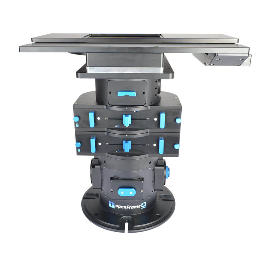
The OpenFrame is a compact, modular, open-source microscope conceived by Paul French and developed with colleagues from the Photonics Group at Imperial College London, and designed, supported, and extended in collaboration with Cairn Research. The fundamental building blocks for self-builders are open source and can be purchased directly from Cairn, or machined in your own workshop based on the STEP files provided on, Photonics Group.
Alternatively Cairn Research can design, build, and test a bespoke microscope for your specific application consisting of any pre-assembled and aligned OpenFrame layers, complete with a choice of light sources, images splitters, stages, cameras, software etc from Cairn or other manufacturers. We are committed to sustainability and will also seek to integrate any components that you may already have in the lab on redundant microscopes to save your hard-earned grant money and to avoid waste.
Total Flexibility – Precision Without Limits.
The OpenFrame Microscope is a next generation imaging platform designed for researchers who demand complete control over their experimental setup. Built around a fully open architecture, it enables seamless integration of advanced components while maintaining exceptional stability and performance.
Whether you’re pushing the boundaries of neuroscience, cell biology, or biophysics, the OpenFrame gives you the freedom to build exactly the system you need without compromise.
Engineered for Freedom
Break free from the constraints of traditional microscope systems. The OpenFrame’s fully open design allows complete customisation, enabling unique optical paths and complex experimental configurations.
Precision You Can Trust
A rigid, high-stability frame minimises vibration and drift, ensuring consistent, high quality imaging even in the most demanding applications.
Built Around Your Workflow
Every research challenge is different. The OpenFrame adapts to yours, supporting a wide range of components including cameras, stages, lasers, and detectors.
Future-Proof by Design
As your research evolves, so can your system. Expand, upgrade, and reconfigure without needing a complete rebuild.
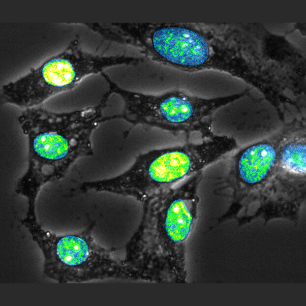
Fluorescence & Contrast Imaging
– Wide-field fluorescence
– Phase contrast
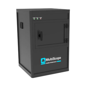
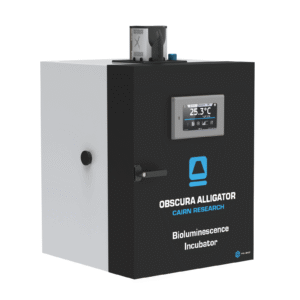
– Bioluminescence
Functional & Quantitative Techniques
– FLIM
– Calcium & voltage imaging
– Polarisation / anisotropy studies
Advanced & Super-Resolution
– STORM, PALM, SIM
– Single-molecule imaging
– TIRF
Dynamic & Interactive Imaging
– FRAP
– Optogenetics
Integrated & Scalable Workflows
– Electrophysiology
– High-content imaging
– SPIM / light-sheet microscopy
Specialised Environments
– Containment labs
– Sterile / easily sterilised setups
The OpenFrame is not just a microscope, it’s a platform for innovation. Designed in collaboration with Cairn Research and Paul French and developed with colleagues from the Photonics Group at Imperial College London. The fundamental building blocks for self-builders are open source and can be purchased directly from Cairn, or machined in your own workshop based on the STEP files provided on, Photonics Group.
Work with our specialists to create a tailored imaging solution that meets your exact research needs.
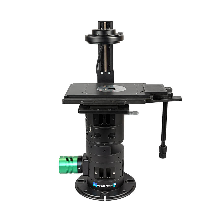
Compact, Stable & Purpose-Built
A space-efficient, stacked modular design delivers exceptional mechanical stability while maintaining full access to the optical path ensuring precision, reproducibility, and ease of use.
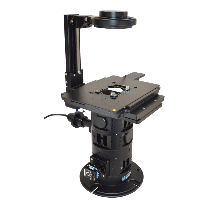
Reconfigurable & Scalable Platform
Quickly adapt the system to new applications with minimal downtime. Start with what you need and expand over time, from routine experiments to complex, high content workflows.
Open, Brand-Agnostic Architecture
Freedom to use components from any manufacturer including Olympus, Nikon, and Thorlabs. Seamlessly integrate existing cameras, stages, illuminators, optics, and filters without vendor lock in.
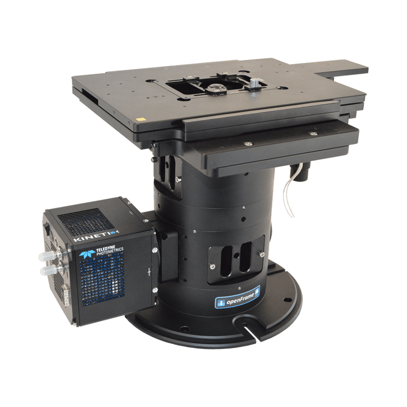
Advanced Imaging Capability
Supports simultaneous multi-camera imaging and large-format sensors (>25mm diagonal), enabling high-throughput data capture and advanced experimental setups.
Built for Innovation & Rapid Development
Provides direct access to the optical train, allowing fast alignment, validation, and prototyping of new techniques. Ideal for developing and testing cutting-edge microscopy methods.
Sustainable & Serviceable
Designed for long-term use, easily maintained, serviced, or modified by any skilled microscopist or technician, reducing reliance on proprietary support.
Flexible Integration & Custom Builds
Available as fully bespoke, turn-key systems tailored to your research needs, or as a modular platform that evolves with your experiments.
Practical for Real-World Use
Easy to clean, autoclavable where required, and transportable, making it suitable for shared labs, teaching environments, tradeshows, and collaborative projects.
Efficient, Future-Proof Investment
A cost-effective entry point that grows with your research, supporting everything from specialised experimental setups to diagnostics and teaching applications.
The OpenFrame Microscope is trusted across a wide range of advanced research fields:
Quantitative evaluation of LED based optical autofocus module – April 2026
Biomolecular condensates sustain pH gradients at equilibrium through charge neutralization – January 2026
Assessing PARP trapping dynamics in ovarian cancer using a CRISPR-engineered FRET biosensor – January 2026
piSTORM: programmable illumination in stochastic optical reconstruction microscopy – November 2025
SlimVar for rapid in vivo single-molecule tracking of chromatin regulators in plants – September 2025
A roadmap for the widespread adoption of frugal microscopes – September 2025
Diffusing protein binders to intrinsically disordered proteins – July 2025
Model‐free machine learning‐based 3D single molecule localisation microscopy – May 2025
Open-source implementation of polarisation-resolved single-shot differential phase contrast microscopy (pDPC) on a modular openFrame-based microscope – March 2025
Fluoro-Electrochemistry Based Phytoplankton Bloom Detection and Enumeration; Field Validation of a New Sensor for Ocean Monitoring – October 2024
A prenatal skin atlas reveals immune regulation of human skin morphogenesis – October 2024
Super-resolved fluorescence imaging utilising accessible stochastic optical reconstruction microscopy (easySTORM) implemented on a low-cost, modular open-source (openFrame) microscope – September 2024
Mechanical Profiling of Biopolymer Condensates through Acoustic Trapping – September 2024
Innovating in a bioimaging core through instrument development – April 2024
Cost-effective, Modular, Research-grade Optical Microscope – January 2024
Controlling crystallisation and dissolution of biogenic CaCO3 via dissolved magnesium cations – January 2024
The Chromatin Regulator HMGA1a Undergoes Phase Separation in the Nucleus – November 2023
High-Throughput Combinatorial Analysis of the Spatiotemporal Dynamics of Nanoscale Lithium Metal Plating – August 2023
Surface patches induce nonspecific binding and phase separation of antibodies – April 2023
The K2: Open-source simultaneous triple-color TIRF microscope for live-cell and single-molecule imaging – March 2023
Surface patches induce nonspecific binding and phase separation of antibodies – February 2023
Open microscopy in the life sciences: quo vadis? – August 2022
The miEye: Bench-top super-resolution microscope with cost-effective equipment – July 2022
Rapid Opto-electrochemical Differentiation of Marine Phytoplankton – April 2022
Surface interaction patches link non-specific binding and phase separation of antibodies – March 2022
Development and Applications in Super Resolution Microscopy – July 2021
Low-cost, sustainable, modular open microscopy and high content analysis – June 2021
Cairns’s design ethos is based on compatibility across a wide range of manufacturers. In the rare case where we do not have a compatible part, we have a custom design team available to provide a solution.
We provide a comprehensive 12-month warranty on all our products.
We aim for a delivery time of 2-4 weeks. However, the specific delivery time will be confirmed at the point of order.
We offer full training and support across our entire product range.
Yes, we have full custom design capabilities and a multidisciplinary team of scientists and engineers available to provide a solution that meets your needs.
Our CellCams are supported in Micro-Manager.