LSPI Single Plane Illuminator Lightsheet Imaging

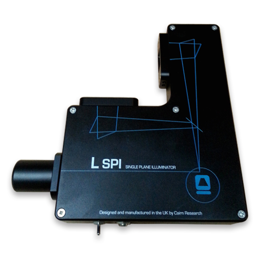
L-SPI Large Selective Plane Illuminator

• Live-cell imaging with ultra-low phototoxicity
• Spatial and temporal fractionation of illumination
• Orthogonal lightsheets reduce shadowing

Product Description

Now complete with X, Y, and Z stages to greatly assist usability 

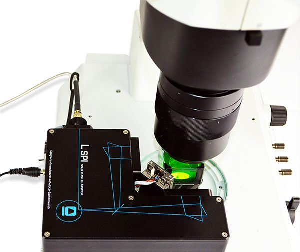
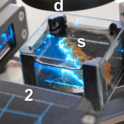
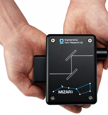
The L-SPI is designed to work with any macro- or microscope, single-mode fibre laser source and scientific camera. This simple and effective instrument recombines two uniform, wide lightsheets at right angles, reducing shadowing without the need for sample rotation or image fusion. The system includes optional sample chambers, fast long travel piezo stage and a second L-SPI head to bring sheets in from four directions. The L-SPI can be adapted to a wide range of experimental purposes. Its compact size and large freedom of access (e.g. for positioning micro-electrodes) make it a highly flexible solution.

Please visit our technical support pages for a write up by our co-designer Philippe Laissue

Download Data Sheet Manuals video support Get a Quote Back to Light Sheet Systems

Additional information

Key Benefits

• Live-cell imaging with ultra-low phototoxicity
• Spatial and temporal fractionation of illumination
• Orthogonal lightsheets reduce shadowing
• Uniform lightsheets for macroscopic observation of large samples down to individual cells
• Precise, fast and long-travel piezo stage
• Easy to use – software and microscope agnostic

Specifications

• Single or dual L-SPI heads generate 2 or 4 orthogonal sheets
• 22um or 11um thick sheets as standard
• 1.6mm confocal range with 22um sheet
• sheet X-Y field of view 16x16mm
• Continuous sweeping of focal plane for reduced power density and improved homogeneity
• Fast precise piezo stage
• 20mm of z travel, 10mm/sec speed, 10nm minimum step size
• Magnetic mounting platform for single or dual L-SPI
• Complementary LaserBank with SM fibres

Applications & Publications

2022

Dyer, L., Parker, A., Paphiti, K., & Sanderson, J. (2022). Lightsheet
microscopy. Current Protocols, 2, e448. doi: 10.1002/cpz1.448

Assessing phototoxicity in live fluorescence imaging
LITE microscopy: A technology for high numerical aperture, low photobleaching fluorescence imaging
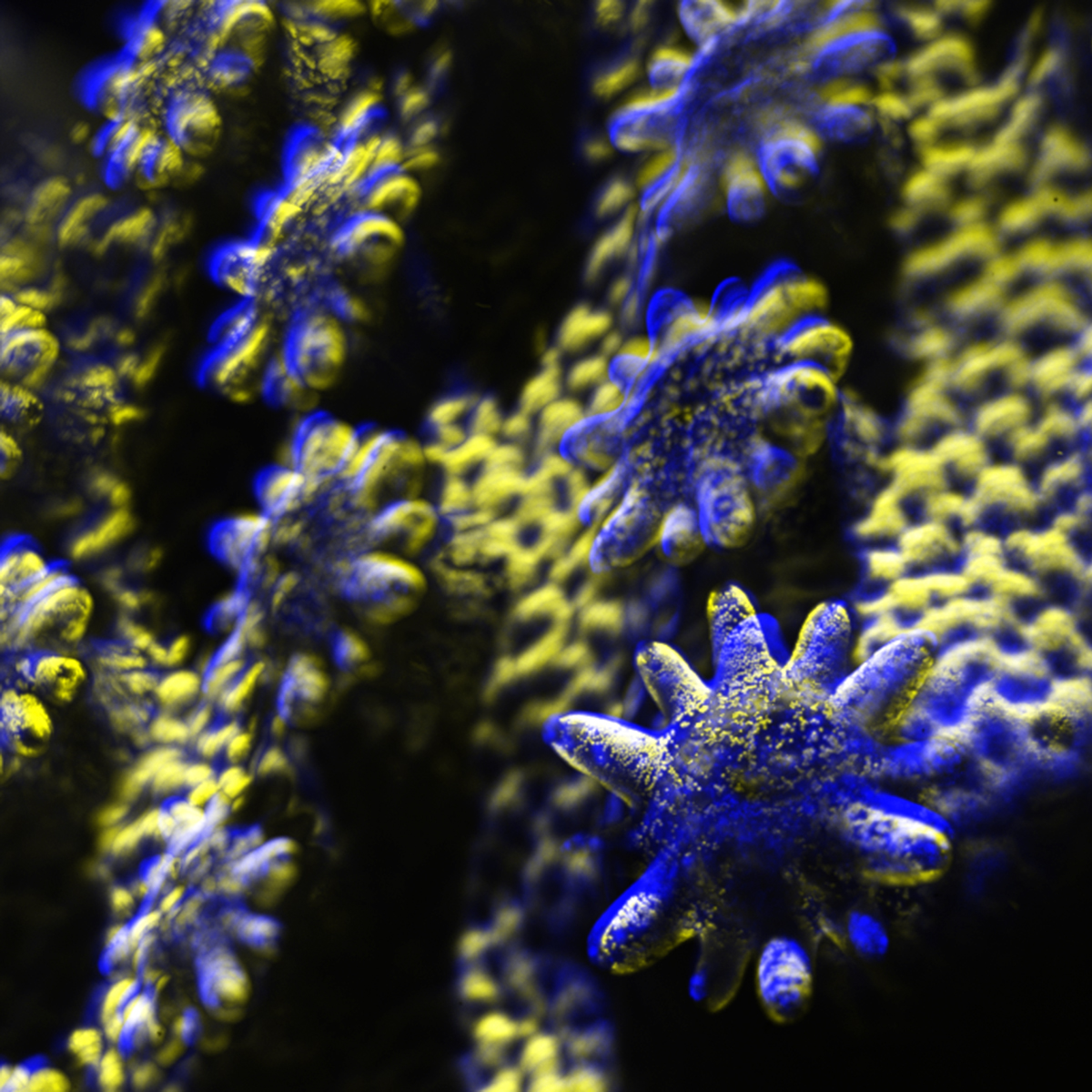
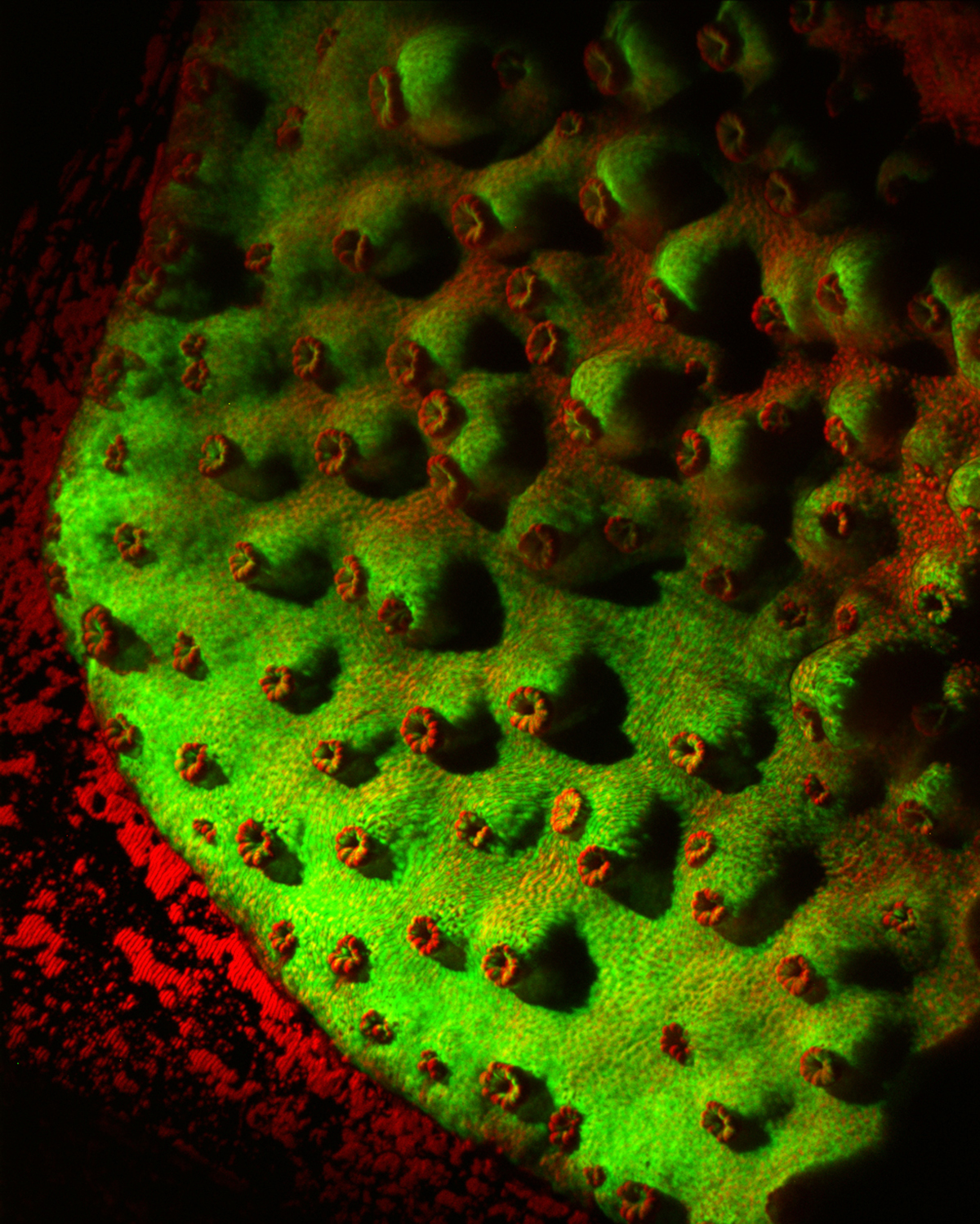
Light sheet microscope tackles live coral imaging


Publications

• McNamara 20171227W draft – smFISH and qIF plus optical clearing Li Germain Gerner 2017 PNAS