Customer Journeys Part 3 – OptoSplit II Simultaneous Dual Colour Capture
In this new series we are highlighting some of the challenges our customers have been faced with and what solutions our technical sales team have provided
![](https://cairn-research.co.uk/wp-content/uploads/2020/03/Richard_Wheeler__System_photo-crop.jpg)
Introduction - Customer Research Challenge
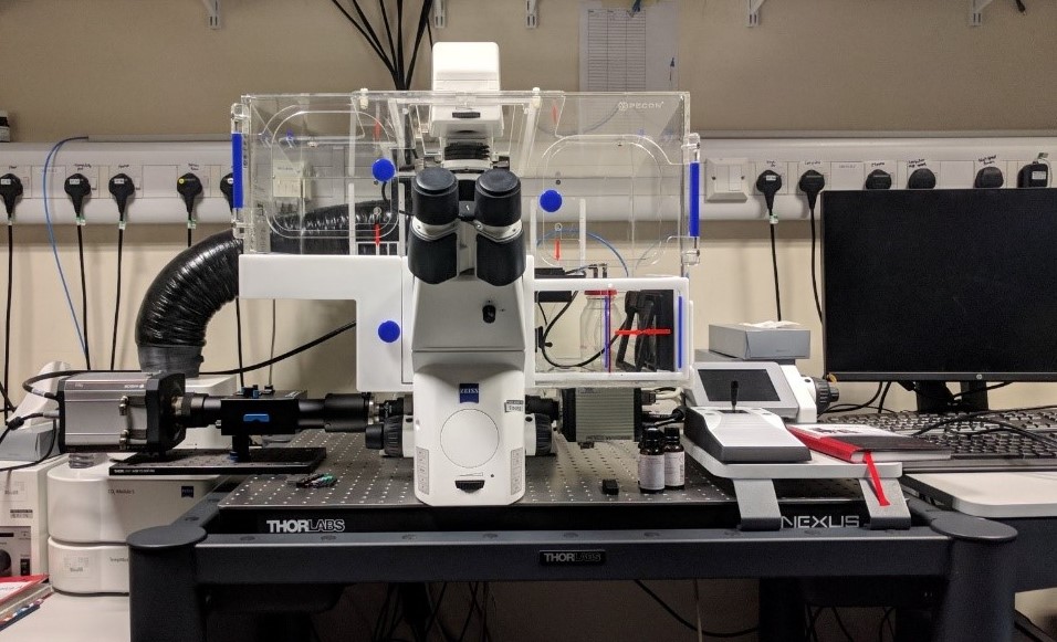
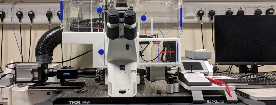
The laboratory of Dr Richard Wheeler studies Leishmania parasites, which are highly motile unicellular eukaryotes; this motility is vital for their life cycle. The unicellular Leishmania parasite is responsible for causing the disease leishmaniasis, which affects millions of people in the developing world. A long-standing goal of the Wheeler group is to be able to visualise the dynamics of intracellular fluorescently tagged proteins during movement, particularly proteins in the flagellum. This presents a challenge due to the high frame rates (>100 frames per second) required to ‘freeze’ flagellar movement combined with the necessity of simultaneous capture of two fluorescent channels to analyse co-localisation.
![](https://cairn-research.co.uk/wp-content/uploads/cmdm/10324/1545228582_OptoSplit-II-Large-300x199.png)
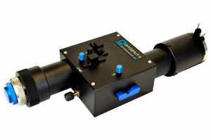
OptoSplit II dual emission image splitter
Traditional approaches to dual wavelength imaging such as a motorised filter changer or an additional camera and beamsplitter are less than ideal in this scenario, as the switching speed of an electronic filter changer limits temporal resolution, whilst a second camera adds cost and complexity to a microscope system.
Cairn Research Solution
The ‘OptoSplit II’ image splitter, when combined with a suitable camera, can enable high frame rate, simultaneous, dual colour imaging by projecting fluorescence from one fluorescently-labelled group on to one half of the camera sensor and that from a second group on to the other half of the sensor. This allows the dynamics of two highly mobile populations of molecules, where each group is labelled with a different fluorescent reporter, to be tracked at the same time on a single camera. In their most recent bioRxiv preprint, the Wheeler group used an OptoSplit II coupled to a Neo sCMOS camera (Andor Technology, UK) to study asymmetries in flagellum beating in moving Leishmania. In one set of experiments they simultaneously monitored at high frame rate a flagellum membrane marker labelled with mCherry and a marker of the asymmetric microtubule-based cytoskeletal structure labelled with mNeonGreen.
![](https://cairn-research.co.uk/wp-content/uploads/2020/03/Richard_Wheeler__System_photo.jpg)
Conclusion
The group of Dr Richard Wheeler is interested in elucidating the processes responsible for flagellum-based motility in the Leishmania parasite and they use a combination of molecular and biochemistry techniques, supported by mathematical and computational simulation, biophysics and automated image analysis to achieve this. They adapted one of their existing inverted microscope frames for dual colour high frame rate widefield epifluorescence microscopy using an OptoSplit II and a sCMOS camera, thereby allowing them to monitor and track multiple fluorescently labelled molecules at a high temporal resolution. This approach has provided new insights into the mechanics of Leishmania flagellum beating, which will potentially aid the development of novel anti-parasitic treatments to combat leishmaniasis.
The OptoSplit II has provided me with a very cost effective and easy-to-use solution to capturing dual colour fluorescence synchronously on a high-speed camera. Its self-contained nature is ideal for cleanliness (for pathogen research) and allows easy reconfiguration of the microscope for other experiments.
Dr Richard Wheeler, University of Oxford