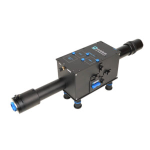
The OptoSplit III (TripleSplit) extends the OptoSplit image splitter concept, adding an optional second beamsplitter to split the field into either two or three separate, spatially equivalent, channels, which can be displayed side by side on a single camera chip.
Splitting is usually performed on the basis of wavelength or polarisation, allowing applications where there is a requirement for simultaneous, or high speed, acquisition of multiple emission bands or polarisations states. The simultaneous acquisition of up to three images offers a major benefit over manual or electronic filter changers, as there is no longer a need to pause acquisition while the filter position is changed. This allows your camera to be operated in fast stream modes.
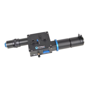
Device drivers are included in most commercial imaging packages to assist registration and to allow realtime and off-line ratioing or fluorescence overlays. Alternatively the TripleSplit can be used with simple image capture software and the processing carried out manually offline or using our own MicroManager and ImageJ drivers. The simple and accessible design makes the Optosplit III an excellent platform for alternative applications, such as pFRET polarisation, or multi-depth imaging.
Whilst optimised for coupling to a scientific microscope, the Optosplit III image splitters can also be used with camera lenses or any other system of lenses which produce an image plane of suitable size and f/number (please ask for details).
SIMple: A fibre-based platform for accessible structured illumination microscopy. bioRxiv (Cold Spring Harbor Laboratory) — September 2025
The role of actin dynamics in vesicle formation during clathrin-mediated endocytosis. bioRxiv (Cold Spring Harbor Laboratory) — August 2025
Imaging the dynamics of vesicle formation supports the flexible model of clathrin-mediated endocytosis. Biophysical Journal — February 2022
TCR-pMHC bond length controls TCR ligand discrimination. The Journal of Immunology — 2019
Short Linear Sequence Motif LxxPTPh Targets Diverse Proteins to Growing Microtubule Ends. Structure — June 2017
Real-time fluorescence imaging with 20 nm axial resolution. Nature Communications — June 2015
A Stochastic Model for Electron Multiplication Charge-Coupled Devices – From Theory to Practice. PLOS ONE — January 2013
Cairns’s design ethos is based on compatibility across a wide range of manufacturers. In the rare case where we do not have a compatible part, we have a custom design team available to provide a solution.
We provide a comprehensive 12-month warranty on all our products.
We aim for a delivery time of 2-4 weeks. However, the specific delivery time will be confirmed at the point of order.
We offer full training and support across our entire product range.
Yes, we have full custom design capabilities and a multidisciplinary team of scientists and engineers available to provide a solution that meets your needs.
Our CellCams are supported in Micro-Manager.